




Microglia and macrophages
Minor accidents caused by everyday activities, as well as acute inflammatory processes triggered by pathogens and the associated discomforts—such as fever, swelling, or pain— are familiar to everyone, since this is the immune system’s primary response even to the slightest injury—but what happens in the brain when there is chronic inflammation somewhere in our body?
The latest article by Ádám Dénes’s group was published in Nature Communications, and it comes as no surprise that the main focus here is once again on microglia—more specifically, microglia and border zone macrophages. These immune cells are located in different areas of the brain, and although we have long known about their distinct functions, their roles during systemic inflammation have not yet been compared.
The study’s first authors, Ana Rita Brás and Nikolett Lénárt, along with Ádám Dénes, who leads both the research and the research team, answer these questions.
- Was it predictable—or not—that chronic inflammation would alter the interactions between microglia and the vascular system?
Rita, Niki, Ádám:
- We knew that microglia constantly monitor the condition of the brain’s blood vessels and that their processes migrate to areas of inflammation or injury. What is less well known, however, is what changes occur in the brain’s blood vessels as a result of systemic inflammation, how this affects the state of the microglia, and what vascular processes the microglia help regulate.
We sought to understand this by developing a model in which repeated peripheral administration of the inflammatory bacterial lipopolysaccharide (LPS) sustains elevated levels of inflammatory factors—which also affect cerebral blood vessels—in the circulation for several days, without any direct physical manipulation of the brain.
- Could you give an example of this occurring within this organization?
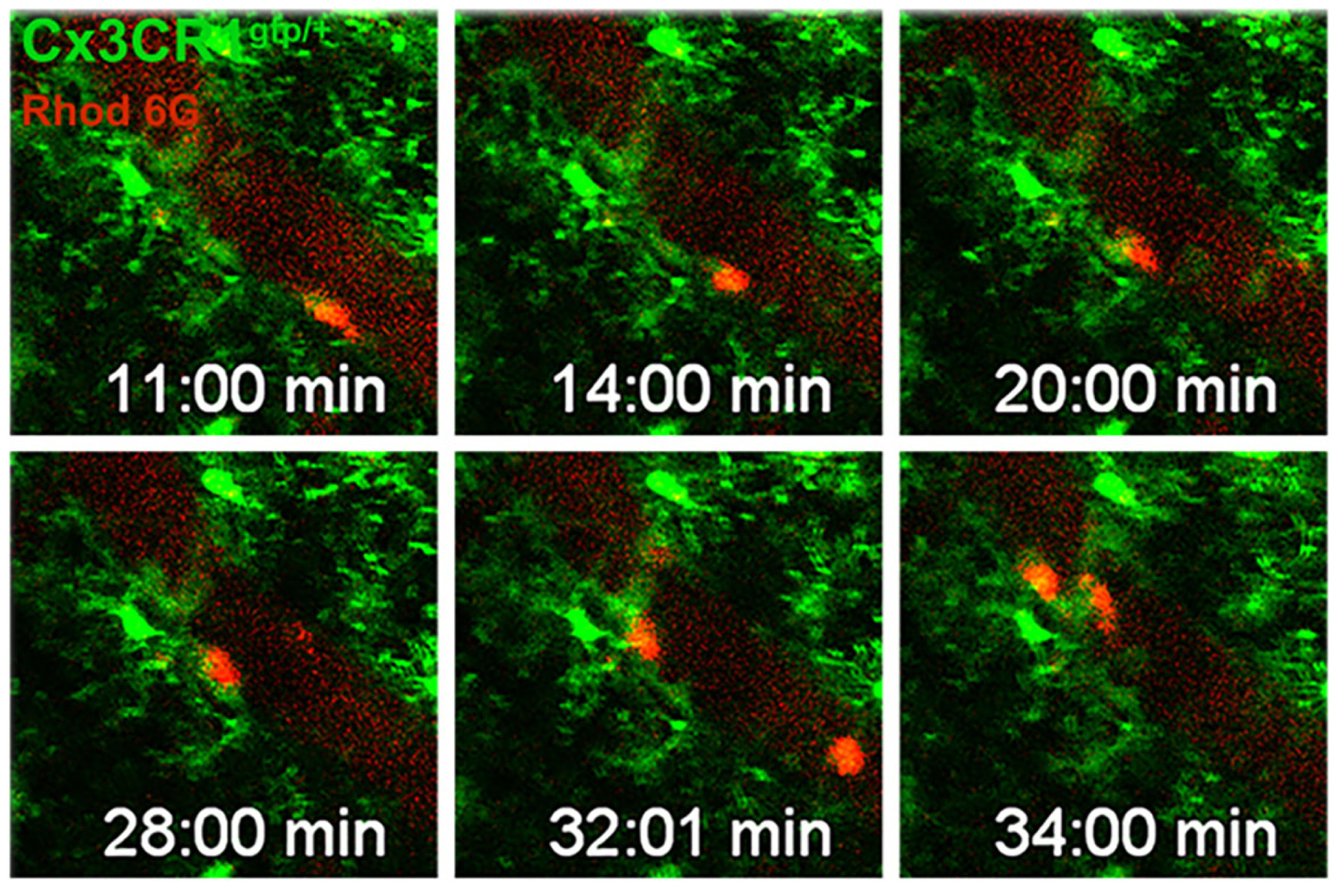
- It’s comparable to the stress our body experiences during an infection lasting several days, such as pneumonia. While it’s certainly not surprising that such prolonged inflammation alters the state of microglia, our model provided an opportunity to investigate microglia-dependent vascular processes during chronic systemic inflammation. We found that, as a result of inflammation, numerous white blood cells were recruited from the circulation to the cerebral blood vessels, and that microglia, as well as so-called border zone macrophages located in the meninges and around cerebral blood vessels, play a role in this process—though they induce changes in opposite directions. While microglia promoted the recruitment of white blood cells, border zone macrophages inhibited it. We made similar observations regarding the role of these cells in cerebral circulation as well. Systemic inflammation impaired cerebral circulation, and this impairment worsened when microglia were absent or dysfunctional, but improved when border zone macrophages were removed.
- What is the relationship between systemic inflammation and changes in cerebral circulation triggered by elevated CO2 levels (hypercapnia)?
- Cerebral circulation is subject to extremely dynamic regulation. Let’s start with a well-known fact: although our brain accounts for only 2 percent of our body weight, it consumes nearly 20 percent of the circulating oxygen and glucose during its functioning. To ensure the uninterrupted functioning of such an energy-intensive system, it is necessary to maintain the independence of cerebral circulation from systemic blood pressure within a certain range —this is what we call autoregulation—as well as neurovascular coupling, which is the coordination of neuronal activity and blood flow to ensure that specific brain regions receive an increased blood supply during periods of heightened activity.
- How is hypercapnia related to this truly remarkable system?
- In our previous work, we demonstrated that microglia can influence both autoregulation and neurovascular coupling by affecting changes in blood vessel diameter. From a physiological standpoint, the key trigger for an increase in blood vessel diameter is elevated carbon dioxide levels (hypercapnia), which leads to increased cerebral blood flow. This is why we do not immediately lose consciousness when we hold our breath.
We know that inflammation impairs the regulation of cerebral circulation, but our recent studies have now confirmed that this is due to changes in the state of the microglia.
- In one part of your experiments, you removed microglia and/or border zone macrophages. The absence of both represents a very significant change compared to the natural state—how should the resulting findings be interpreted?
- We employed various cell manipulation methods to map the selective effects of different brain macrophage/microglia populations. In another series of experiments, we removed interleukin-1 (IL-1), one of the most important inflammatory cytokines, from the microglia, and this also resulted in reduced white blood cell recruitment. In addition, we discovered that the two IL-1 isoforms influence cell recruitment and cerebral circulation differently: the removal of microglial IL-1α and IL-1β reduced the number of leukocytes, while inhibition of IL-1α improved cerebral circulation. It also turned out that the effects of vascular inflammation on arterial and venous segments depend on IL-1 in different ways, and the connections between macrophages/microglia in the border zone and blood vessels are not the same for the respective vascular segments!
- This system is not only amazing but also astonishingly complex! Can we determine the proportions of different macrophage populations in the brain and how they differ from our favorite microglia?
- With the widespread adoption of modern “cell fate mapping” (fate-mapping) methods and single-cell sequencing, it has now become clear that the overwhelming majority of both microglia and border zone macrophages originate from the extraembryonic sac and form self-sustaining populations in various regions of the brain from a very early stage of ontogenesis. The name “zone of interface macrophages” derives from the fact that these cells are found in the meninges, the walls of blood vessels, and the choroid plexus—areas where protective brain structures, or “barriers,” also form, isolating the brain’s microenvironment from the peripheral circulation: blood-brain barrier, blood-CSF barrier, meningeal barrier structures, etc.). The only “macrophages” in the brain parenchyma are the microglia, which account for 5–8 percent of all brain cells—meaning they are present in far greater numbers than border zone macrophages. Their roles also differ: macrophages in the border zone act as “gatekeepers” for barrier structures that are important from an immunological perspective and are vulnerable to damage, while microglia perform numerous physiological functions within the brain tissue through their connections with neurons, blood vessels, and glial cells—such as regulating neuronal activity, facilitating myelination, and protecting cerebral blood vessels.
- What is the most important message—the main point—or lesson of this work, and in what ways is it forward-looking?
- First, it reveals the distinct roles of microglia and border zone macrophages in systemic inflammation, including their roles in the recruitment of white blood cells to the brain and in the regulation of cerebral circulation.
The model used is simple and allows for the mapping of cell-cell interactions and molecular mechanisms. These mechanisms may also be important in brain diseases such as stroke or multiple sclerosis (MS), where the influx of white blood cells into the brain influences the state of blood vessels and neurons in complex ways.
It may also explain why cerebral circulation and the network activity of neurons change during respiratory diseases such as influenza or COVID-19.
Last but not least, our findings may even help us understand the mechanisms through which systemic inflammation affects the condition of cerebral blood vessels and microglia—for example, in conditions like diabetes or atherosclerosis—which subject the body and the brain’s vascular network to a gradual inflammatory burden.
- My compliments to all of you, as your findings are important for several areas of research!
My final question is for Ádám, as the principal investigator: Who and what would you highlight regarding this work?
Ádám:
- Rita and Niki put a tremendous amount of work into preparing this material, and on top of that, they ended up with a reviewer who was clearly personally opposed to the article’s publication. Fortunately, the journal’s editor handled the review process with great patience, professionalism, and impartiality, and after receiving expert opinions from five reviewers, the article was finally accepted.
As the Latin saying goes, per aspera ad astra—the road to the stars is rocky!
In vivo two-photon imaging in the mouse cerebral cortex. Motile microglial processes interact with leukocytes recruited to cerebral blood vessels under systemic inflammatory conditions.