





When the Memory Brings Fear Once Again
No researcher need fear that one day there will be nothing left to discover.
Our understanding of the brain’s anatomy is not yet complete, even though its first comprehensive scientific map—the Brodmann map, which remains well-known today—was published as early as 1909; a hundred years later, in 2011, the Allen Institute for Brain Science published the first genetic map of the human brain, and since 2013, anyone has been able to explore a three-dimensional digital model of the human brain with a resolution of 20 micrometers, and with the help of modern imaging techniques and cell-level analyses, there is still a chance today for anyone who asks the right questions, possesses excellent knowledge of neuroanatomy and methodology, and is not afraid of long-term work to discover previously unidentified clusters of neurons—known in neuroanatomy as “nuclei.”
Gábor Nyiri and his team have already discovered a brainstem region that plays a key role in processing emotions and regulating motivational processes; in their paper just published in Nature Communications, they describe a neural circuit that links memory recall to the fear response.
Here are the answers to our questions from Boldizsár Balog, a Ph.D. student and the first author of the study—answers that reveal not fear, but a thorough understanding of the work!
- Why wasn’t the classical cortical memory system sufficient, and how is the current one different?
- Let’s take the well-known story. Under the yellowish glow of a streetlamp, a man is rummaging around, and his friend, walking by, crouches down beside him to help him find his apartment key. After searching the cold asphalt for a long time to no avail, the friend asks, “Did you see which way it might have bounced?” The man points toward the distant, dark doorway: “I dropped it right there in the doorway, but the lighting is much better here!”
Many people believe that this is exactly how our lives—and brain research—work: we focus on whatever task we’re currently able to solve. The cerebral cortex is easily accessible, so we’re not going to grope around in the dark trying to examine subcortical areas that are difficult to study. However, especially over the last two decades, optogenetics—thanks to the work of Karl Deisseroth, Peter Hegemann, Georg Nagel, and a few others—the development of optogenetics, fiber-optic imaging, electrophysiology, and many other methods has “replaced the lightbulb above the gate,” making even deeper brain regions accessible.
In the case of optogenetics, it has become clear—in more than one sense—that, in addition to the cerebral cortex on the surface, many deeper brainstem structures are also important for memory. The classical model examines the interactions among the prefrontal cortex, hippocampus, and amygdala, but with modern methods, we find that there are plenty of phenomena that cannot be explained by this model.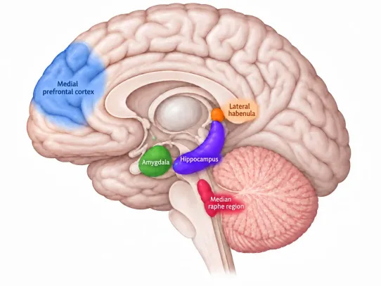
- Let’s move on to the models. What is it that has no equivalent in the rodent model?
- When one of our predecessors pondered why humans rose to the top of the animal kingdom, I don’t think they were thinking of our exceptional physical strength, but rather our intellect. And if that ancestor happened to have some knowledge of neuroanatomy, he would have immediately seen that the human cerebral cortex, for example, is enormous compared to that of a mouse. Our hero might thus have concluded that thinking, self-awareness, and everything else necessary for this dominance must be found in the cerebral cortex. I think he was wrong. Ever since the “lightbulb moment,” neuroscientists have identified areas beneath the cortex that are responsible for empathy, learning, and adaptation. That’s why I think it would be difficult to find anything that doesn’t have at least a simplified counterpart in rodents.
- What are the key cells involved in the recall of a memory and the emergence of the associated fear? How sensitive are these to the treatment used?
- We see that cortical and subcortical areas have evolved together, are closely interconnected, and these connections are also crucial. For example, when we startled mice and the next day, as they recalled the frightening situation, we blocked the neurons projecting from the prefrontal cortex to the median raphe of the brainstem, the mice were much less fearful and recalled the previous day’s negative experience to a lesser extent. In fact, we also observed that they relearned more quickly that they no longer needed to be afraid in this environment. (This is what we call memory extinction.) In short, the cells involved in this process are quite sensitive to treatment.
- The cellular-level explanation is a fantastic finding, but to what extent can we actually target only the relevant cells at the cellular level during treatment—specifically, the brainstem cells involved in anxiety, PTSD, and depression—and how precisely can we treat them?
- The aforementioned process of re-learning is particularly important because we could utilize it, for example, to extinguish fear memories that are easily triggered—a characteristic of PTSD. To prepare for this, in another experiment conducted jointly with KOKI and a Swiss laboratory, we also examined a population of neurons in the aforementioned median raphe region. We found that these cells express relatively specific ion channel and neurotransmitter receptor genes that could potentially be targeted with drugs in the future.
- Do ketamine and other agents already in use act solely on cells that are sensitive to chronic stress? To what extent can a treatment (whether this one or one involving chemical agents) be “fine-tuned”?
- We do not know the exact mechanism of action for most of the drugs used in clinical practice. If a substance has been tested by humanity, has been shown to work in certain situations, and has also been found to be safe, then we use it as a drug. Of course, in modern medicine, very strict regulations must be followed, and a substance must undergo numerous efficacy and safety tests before it can be approved as a drug. We’ve only known for about two years that ketamine targets the lateral habanula—one of the best-known centers of depression—relatively specifically, but it’s no coincidence that it’s also used in clinics in the U.S. to treat depression. In our recently published article, we showed that since the cells in the median raphe that we studied are activated by stress, we can use ketamine to reduce and fine-tune this overactivation.
- What do you consider to be the greatest risk of mood-regulating treatments?
- Perhaps the side effects. Precisely because ketamine, for example, targets NMDA receptors found in so many areas of the brain, it has a wide variety of effects—and thus side effects as well.
In one of our experiments, we used special vectors to introduce an artificial receptor into the median raphe cell population of stressed mice, which allowed us to restore their motivation. Many believe that in the future, such gene therapy methods will enable us to treat patients in a more targeted manner with fewer side effects.
- The use of DBS (e.g., in patients with Parkinson’s disease) has been very helpful in overcoming movement disorders and depression, but in many cases it has apparently altered the patients’ personalities as well. Do we already know the reason for this?
- DBS, or deep brain stimulation, is a non-selective method; within a given area, it can simultaneously affect many different cell types and neural pathways, which is precisely why we can expect various side effects, including personality changes. Of course, in many cases, even with these side effects, we can achieve a much better quality of life compared to the untreated state, so it’s worth using or trying if necessary.
- Is there any insight into what drives patients with depression, PTSD, etc., to relive their fears over and over again? If patients’ brain activity were monitored for days on end, could the process be identified at the cellular level, or are these precisely the specific brainstem cells that, when inhibited, could reduce depression?
- Repetition is the mother of learning, and the neurological basis for this lies in the connections between neurons. For example, if a neuron is very active, its connections with other areas of the brain may strengthen. We observed a similar process in the cells of the median raphe: as a mouse learned that a certain sound signaled a frightening experience, the activity of the cells in the median raphe also increased. Unfortunately, for now, these precise measurements require brain surgery, so we cannot study these processes in human experiments; we have only been able to confirm the presence of our cell population in primates.
I am confident that, within my lifetime, a method will be developed that will allow us to study these cell populations in our own brains and even modulate their function.